|
||||||||||
Insertion of Suprapubic Catheter Retropubic
Urethropexy: Ureteroneocystostomy
and Transperitoneal Intestinal
Loop |
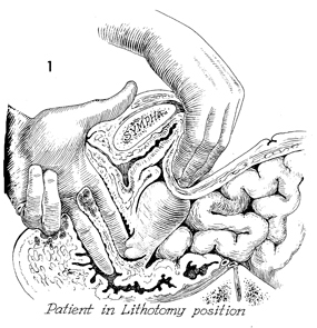
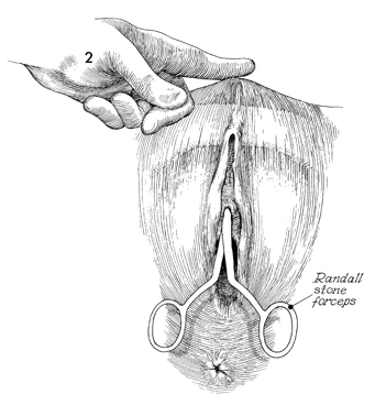
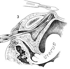
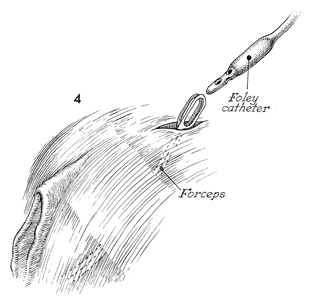
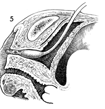
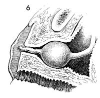
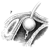
Insertion of Suprapubic Catheter Dissection at the base of the bladder to reach the anterior vaginal wall and uterine cervix creates edema, interrupts the small nerve pathways, and thereby sets up the physiologic changes that produce urinary bladder atony. Therefore, catheter drainage of the urinary bladder is an essential feature of many pelvic surgical procedures. Fortunately, in most cases, these conditions reverse themselves in 3-5 days, and catheter drainage is no longer needed. Suprapubic bladder catheterization is superior to transurethral bladder catheterization because it is cleaner. It also leaves the urethra open for voiding when urinary function has returned. The use of an ordinary Foley catheter (No. 16 French with 5-mL bag) is preferable to the commercially available suprapubic catheter kits because a Foley catheter, when inserted as described in this section, is usually not dislodged from the bladder during sleep or activity. In addition, the Foley catheter is less costly and is available in all surgical clinics. The instrument used for insertion of the Foley catheter is an ordinary Randall stone forceps. The fulcrum of this instrument is toward the rear, which keeps the overall diameter of the axis virtually unchanged except at the jaws and gives it an advantage over a Kelly clamp. The operation provides drainage of the urinary bladder through a clean surgical incision and ensures that the catheter does not slip out of the patient or become dislodged within the abdominal wall. Physiologic Changes. The procedure reduces edema at the base of the bladder, allowing the return of normal vesical function. Points of Caution. After grasping the catheter with the jaws of the Randall forceps (Fig. 4) and before inflating the Foley balloon, the catheter should be drawn through the bladder until the tip can be seen in the urethral meatus. This ensures that the catheter tip and balloon are in the bladder and not in the subcutaneous or subfascial space. Technique
|
|||||||||
Copyright - all rights reserved / Clifford R. Wheeless,
Jr., M.D. and Marcella L. Roenneburg, M.D.
All contents of this web site are copywrite protected.

