Home / Site Map / Vulva and Introitus / Vagina and Urethra / Bladder and Ureter / Cervix / Uterus
Fallopian Tubes and Ovaries / Colon / Small Bowel / Abdominal Wall / Malignant Disease: Special Procedures

Bladder and Ureter

Insertion of Suprapubic Catheter

Retropubic Urethropexy:
Marshall-Marchetti-Krantz
and Burch Operations

Ureteroureterostomy

Ureteroneocystostomy
and Ureteroneocystostomy
With Bladder Flap

Transperitoneal
Ureteroureterostom
End-to-Side Anastomosis

Intestinal Loop
Urinary Diversion

Percutaneous
Nephropyelostomy

Ureteroileoneocystostomy

Clam Gastrocystoplasty

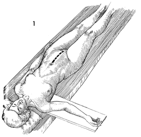
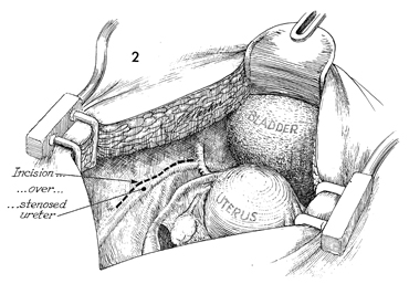
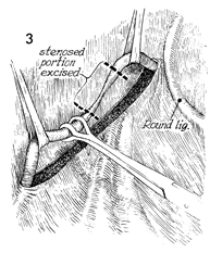
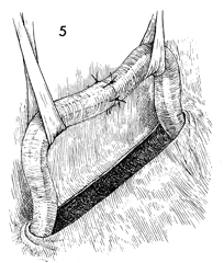
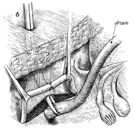
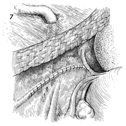
Ureteroureterostomy

Injury to the ureter is occasionally high enough in the pelvis for a primary ureteroureterostomy to be performed without having to resort to a ureteroneocystostomy. In these cases, the ureter has been damaged at or near the pelvic brim while clamping, incising, and ligating the infundibulopelvic ligament or while excising an extensive ovarian carcinoma that has distorted the pelvic anatomy at or near the pelvic brim. Although less common than procedures for correcting lower injuries to the ureter, a ureteroureterostomy is preferable to a ureteroneocystostomy when it can be performed without tension on the anastomosis producing the stenosis.

The essential features of the procedure are the adequate mobilization of the cut ends of the ureter to prevent tension on the anastomosis, the use of a spatulated anastomosis, and the use of delicate suture, meticulous hemostasis, and drainage of the anastomosis site via a closed suction drain through the lower abdominal wall.

The purpose of the ureteroureterostomy is to anastomose the transected ureter.

Physiologic Changes.  After a damaged or diseased portion of the ureter has been removed, the ureter is anastomosed. The sequelae of ureteral obstruction and/or laceration are relieved.

Points of Caution. Care should be taken to see that 
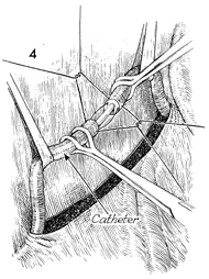
the ureter is anastomosed without tension.

A soft Silastic indwelling catheter should be placed through the anastomotic area and fed into the bladder caudad and the renal pelvis cephalad.

The drain should be placed in the area of the anastomosis and brought out through the right or left lower quadrant and kept in place until all external drainage has ceased.

Technique

The patient is placed in the dorsal position, and the abdomen is opened through a lower midline incision.

The pelvis is cleared of adhesions and intestinal contents. Exposure of the pelvic structures is essential at all times. The pathologic site in the ureter is identified, and the peritoneum overlying the ureter at its junction with the common iliac artery is incised. Dissection of the ureter is carried down to the site of damage and/or stenosis.

An appropriate segment of ureter is dissected out of its bed and mobilized between soft Silastic drains. Care is taken not to damage the ureteral sheath or the delicate network of vessels beneath the sheath that are vital to the vascularity of the ureter. The pathologic portion of ureter is excised with scissors.

A Silastic ureteral catheter is inserted up to the renal pelvis and down into the bladder. Interrupted 4-0 synthetic absorbable sutures are placed through the entire wall of the ureter.

The anastomosis has been completed over a Silastic ureteral catheter. Vessel loops are shown elevating the ureter.

A closed suction drain site in the lower quadrant of the abdomen is selected. A Kelly clamp is passed retroperitoneally, and a soft closed suction drain is brought out through the lower quadrant and is left adjacent to the anastomosis. The drain is used to prevent the collection of urine in the area of the anastomosis and should be left in place until all urinary drainage through it has ceased.

The peritoneum is closed with interrupted 3-0 synthetic absorbable sutures over the ureteroureterostomy so that the ureter remains retroperitoneal.

The Silastic ureteral catheter is removed at the time of water cystoscopy 10-12 days postoperatively. A urologic workup should be performed 6 weeks following anastomosis and 3 months thereafter to ensure against stenosis and hydronephrosis.


Copyright - all rights reserved / Clifford R. Wheeless, Jr., M.D. and Marcella L. Roenneburg, M.D.
All contents of this web site are copywrite protected.