Home / Site Map / Vulva and Introitus / Vagina and Urethra / Bladder and Ureter / Cervix / Uterus
Fallopian Tubes and Ovaries / Colon / Small Bowel / Abdominal Wall / Malignant Disease: Special Procedures

Fallopian Tubes
and Ovaries

Laparoscopy Technique

Diagnostic Uses
of Laparoscopy

Demonstration
of Tubal Patency
via Laparoscopy

Laparoscopic Resection
of Unruptured
Ectopic Pregnancy

Ovarian Biopsy
via Laparoscopy

Electrocoagulation of
Endometriosis via
Laparoscopy

Lysis or Adhesions
via Laparoscopy

Control of Hemorrhage
During Laparoscopy

Fallopian Tube
Sterilization

Sterilization by
Electrocoagulation and
Division via Laparoscopy

Silastic Band Sterilization
via Laparoscopy

Hulka Clip Sterilization
via Laparoscopy

Sterilization by the
Pomeroy Operation

Sterilization by the
Modified Irving Technique

Sterilization by the
Minilaparotomy Technique

Sterilization - Ucheda Technique

Salpingectomy

Salpingo-oophorectomy

Fimbrioplasy

Tuboplasty -
Microresection
and Anastomosis
of the Fallopian Tube

Wedge Resection
of the Ovary

Torsion of the Ovary

Ovarian Cystectomy

Sterilization by the
Minilaparotomy Technique

Minilaparotomy is ideal for thin women with no pelvic disease or adhesions. The procedure is difficult to perform in obese women or in women who have had inflammatory disease of the Fallopian tubes.

In thin, small patients it has the advantage of being performed with instruments less costly than those for laparoscopy. When the patients are given a choice, however, they usually prefer laparoscopy because recovery is faster and less painful and they can 
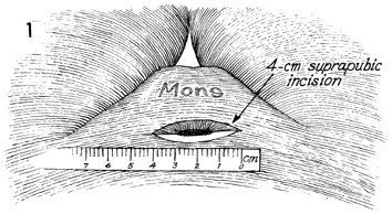
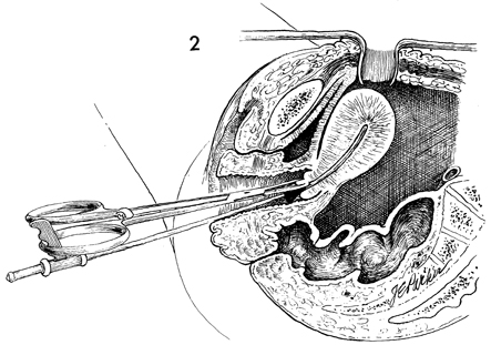
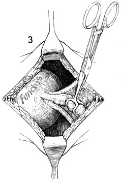
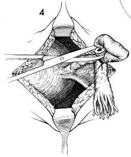
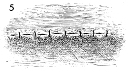
resume their activities much sooner.

The purpose of the procedure is to obstruct the Fallopian tubes.

Physiologic Changes. The Fallopian tubes are obstructed.

Points of Caution. The bladder must be empty, or cystotomy can result. If more than 4 cm are needed to enter the abdomen - the width of 2 adult fingers - the patient is too obese for this operation, and a laparotomy should be performed with the patient under general anesthesia.

Technique

The patient is placed in the dorsal lithotomy position, and a thorough examination of the pelvis is performed to rule out the presence of adnexal disease. The vagina is surgically prepped. A Rubin cannula and Jacobs tenaculum are inserted into the cervix and through the cervical os, respectively. The abdomen is opened with a 4-cm transverse incision above the mons pubis.

A small self-retaining retractor is inserted through the abdominal wall into the peritoneal cavity. The surgeon manipulates the previously placed Rubin cannula and Jacobs tenaculum on the cervix so that the fundus and cornua become readily visible through the small abdominal incision.

A Babcock clamp is used to reach through the incision and grasp the Fallopian tube.

The Fallopian tube is pulled up, and a piece of 0 synthetic absorbable suture is placed around a knuckle of the tube, which is excised with scissors.

The Rubin cannula is manipulated to the other side, and a similar procedure is performed on the opposite Fallopian tube.

The abdominal wall incision is closed in layers. The skin can be closed with either subcuticular or through-and-through sutures.


Copyright - all rights reserved / Clifford R. Wheeless, Jr., M.D. and Marcella L. Roenneburg, M.D.
All contents of this web site are copywrite protected.