|
||||||
Directed Biopsy of the Cervix at Colposcopy Endocervical
Curettage Conization
of the Abdominal
Excision Correction
of an Incompetent Cervix Correction
of an Incompetent Cervix |
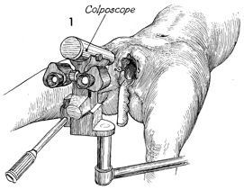
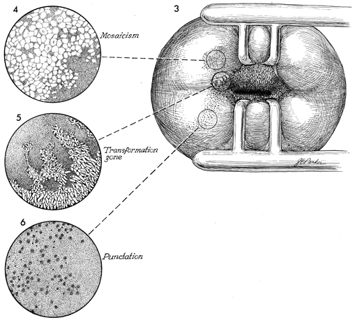
Directed Biopsy of the Cervix Colposcopy as an adjunctive diagnostic tool in the assessment of cervical intraepithelial neoplasia is a significant aid to the pelvic surgeon in selecting the appropriate method of therapy in certain cases. Its use is indicated in all patients having abnormal Papanicolaou cytologic smears or gross lesions. To obtain accurate cytologic specimens for study, the surgeon must be trained not only in performing a colposcopy but also in selecting the proper instruments for the examination. The purpose of the operation is to visualize the cervix under high magnification and delineate abnormal zones of cervical epithelium. In addition, it is designed to allow precise, accurate biopsies to be obtained from these abnormal zones of epithelium. Physiologic Changes. None. Points of Caution. A direct Papanicolaou smear should
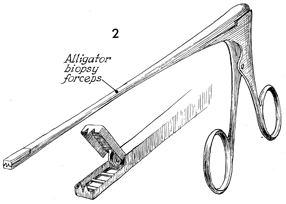
be taken prior to any manipulation of the cervix. A detailed survey of the cervix should be performed prior to any surgical manipulation. Directed biopsies, oriented to prevent tangential cutting, should be placed on small pieces of wet paper towel and sent to the pathologist. Technique
|
|||||

Copyright - all rights reserved / Clifford R. Wheeless,
Jr., M.D. and Marcella L. Roenneburg, M.D.
All contents of this web site are copywrite protected.