Home / Site Map / Vulva and Introitus / Vagina and Urethra / Bladder and Ureter / Cervix / Uterus
Fallopian Tubes and Ovaries / Colon / Small Bowel / Abdominal Wall / Malignant Disease: Special Procedures

Cervix

Biopsy of the Cervix

Directed Biopsy of the Cervix at Colposcopy

Endocervical Curettage
at Colposcopy

Conization of the
Cervix by the Loop Electrical Excision Procedure (LEEP)

Cryosurgery of Cervix

Conization of Cervix

Abdominal Excision
of the Cervix Stump

Correction of an Incompetent Cervix
by the Shirodkar
Technique

Correction of an Incompetent Cervix
by the McDonald
Operation

Correction of an Incompetent Cervix
by the Lash Operation

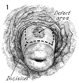
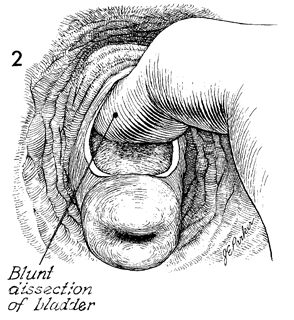
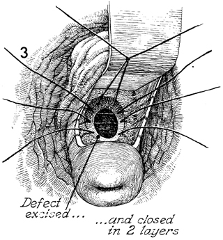
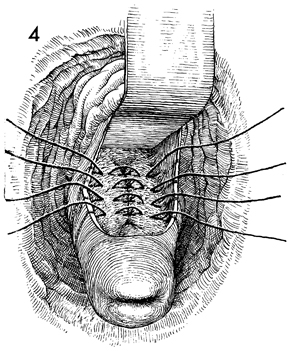
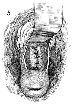
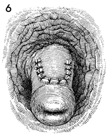
Correction of an Incompetent Cervix by the Lash Operation

Patients with habitual second-trimester abortions occasionally show a defect in the wall of the cervix. This is usually at the 12 o'clock position at the internal cervical os and may be related to failure of fusion of the Mullerian duct. In any case, it results in a weakened internal cervical os. Repair is best performed in the nonpregnant state.

The purpose of the Lash operation is to correct the defect in the cervical wall and restore competence to the cervix, thereby allowing gestation to proceed to term.

Physiologic Changes.  The cervix regains the strength to hold the developing fetus in utero until term, thus preventing second-trimester abortion.

Points of Caution. Patients undergoing the Lash operation should be delivered at term by cesarean section. Dilation and effacement of the cervix following this procedure are unpredictable.

Care must be taken to ensure that the bladder is properly retracted in order to prevent the placement of sutures through the bladder in the closing of the cervical defect.

Technique

The patient is placed in the dorsal lithotomy position under general anesthesia, and the vulva and vagina are prepped in a surgical manner. A weighted posterior retractor is placed in the vagina. The cervix is exposed and is placed on traction with a wide-mouthed tenaculum such as a Jacobs tenaculum. A uterine sound is placed in the cervical os, and the defect is demonstrated in the cervical wall. A transverse vaginal incision is made for approximately 2-3 cm at the junction of the vaginal mucosa and the portio of the cervix. This may require extending the mucosal incision laterally in a "block U."

The bladder is dissected off the cervix with blunt dissection.

The defect is exposed, and the unhealthy cervical tissue is excised with a knife. The opening is closed in two layers with interrupted 2-0 Dexon suture.

The bladder is elevated by a right-angle retractor to separate it from the suturing procedure. Interrupted 2-0 delayed absorbable Lembert inverting sutures are placed in a second row.

The sutures are tied, inverting the defect into the cervical canal. A uterine sound is passed through the cervical canal to ensure that the canal has not been obliterated.

The vaginal mucosa is reapproximated with interrupted 3-0 synthetic absorbable sutures. The patient should be protected from pregnancy for minimum of 3 months to allow maximum healing of the cervical incision.


Copyright - all rights reserved / Clifford R. Wheeless, Jr., M.D. and Marcella L. Roenneburg, M.D.
All contents of this web site are copywrite protected.