Home / Site Map / Vulva and Introitus / Vagina and Urethra / Bladder and Ureter / Cervix / Uterus
Fallopian Tubes and Ovaries / Colon / Small Bowel / Abdominal Wall / Malignant Disease: Special Procedures

Cervix

Biopsy of the Cervix

Directed Biopsy of the Cervix at Colposcopy

Endocervical Curettage
at Colposcopy

Conization of the
Cervix by the Loop Electrical Excision Procedure (LEEP)

Cryosurgery of Cervix

Conization of Cervix

Abdominal Excision
of the Cervix Stump

Correction of an Incompetent Cervix
by the Shirodkar
Technique

Correction of an Incompetent Cervix
by the McDonald
Operation

Correction of an Incompetent Cervix
by the Lash Operation

Endocervical Curettage at Colposcopy

Colposcopy as an adjunctive diagnostic tool in the assessment of cervical intraepithelial neoplasia is a significant aid to the pelvic surgeon in selecting the appropriate method of therapy in certain cases. Its use is indicated in all patients having abnormal Papanicolaou cytologic smears or gross lesions.

To obtain accurate cytologic specimens for study, the surgeon must be trained not only in performing a colposcopy but also in selecting the proper instruments for the examination.

The purpose of the operation is to visualize the cervix under high magnification and delineate abnormal zones of cervical epithelium.

Endocervical curettage enables the surgeon to take specimens from the endocervical canal that may not be visible even with the colposcope.

Physiologic Changes. None.

Points of Caution. A direct Papanicolaou smear should be taken prior to any manipulation of the cervix.

A detailed survey of the cervix should be performed prior to any surgical manipulation.
The endocervical curettings should be sent as a second specimen.

Technique

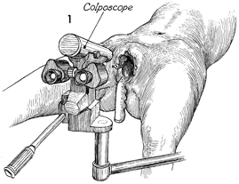
The patient is placed on an ordinary gynecologic examination table, and a nonlubricated speculum is placed in the vagina. The colposcope is moved into appropriate position and focused. A routine cytologic smear as well as a wet mounted smear for Trichomonas and Monilia are taken. The cervical mucus is then cleared by applying 4% acetic acid to the cervix and removing the solution with either a small suction cannula or a cotton-tipped applicator. The cervix is studied carefully with the green filter lens in place to enhance the appearance of the cervical blood vessels.

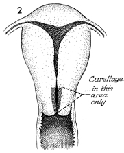
A sagittal section of the uterus and upper vagina shows the area of the endocervix to be curetted. The cervix should not be dilated, since dilation would increase the possibility of the curet entering the endometrial cavity.

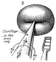
The endocervical curet is placed in the cervix up to the internal os. The curet is moved back and forth in the cervical canal, collecting tissue within the rectangular box. The tissue is then sent to the pathologist. This movement is repeated in a 360° circle until the entire cervical canal has been curetted.


Copyright - all rights reserved / Clifford R. Wheeless, Jr., M.D. and Marcella L. Roenneburg, M.D.
All contents of this web site are copywrite protected.