Home / Site Map / Vulva and Introitus / Vagina and Urethra / Bladder and Ureter / Cervix / Uterus
Fallopian Tubes and Ovaries / Colon / Small Bowel / Abdominal Wall / Malignant Disease: Special Procedures

Vagina and Urethra

Anterior Repair and Kelly Plication
           
Site Specific Posterior Repair
           
Sacrospinous Ligament Suspension of the Vagina
           
Vaginal Repair of Enterocele
           
Vaginal Evisceration
           
Excision of Transverse Vaginal Septum
           
Correction of Double-Barreled Vagina
           
Incision and Drainage of Pelvic Abscess via the Vaginal Route
           
Sacral Colpoplexy
           
Le Fort Operation
           
Vesicovaginal Fistula Repair
           
Transposition of Island Skin Flap for Repair of Vesicovaginal Fistula
           
McIndoe Vaginoplasty for Neovagina
           
Rectovaginal Fistula Repair
           
Reconstruction of the Urethra
           
Marsupialization of a Suburethral Diverticulum by the Spence Operation
           
Suburethral Diverticulum via the Double-Breasted Closure Technique           
           
Urethrovaginal Fistula Repair via the Double-Breasted Closure Technique
           
Goebell-Stoeckel Fascia Lata Sling Operation for Urinary Incontinence
           
Transection of Goebell-Stoeckel Fascia Strap
           
Rectovaginal Fistula Repair via Musset-Poitout-Noble Perineotomy

Sigmoid Neovagina

Watkins Interposition Operation

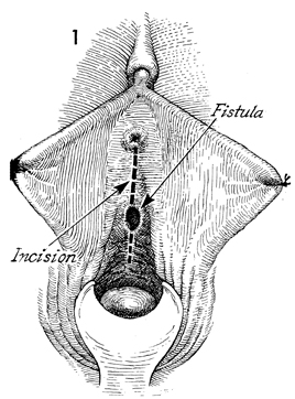
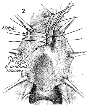
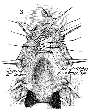
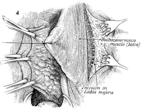
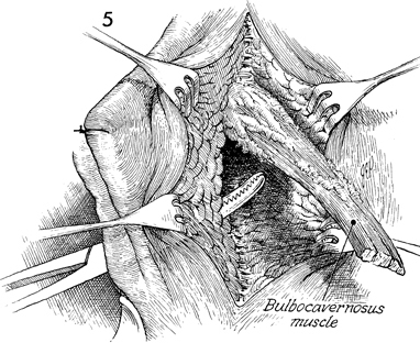
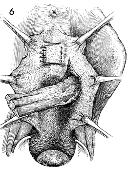
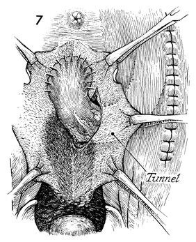
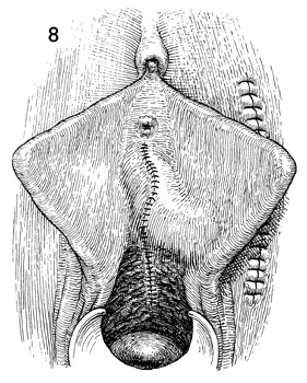
Urethrovaginal Fistula Repair via the Double-Breasted Closure Technique 

Urethrovaginal fistulae are generally secondary to one of two conditions: (1) surgical trauma following anterior colporrhaphy or (2) obstetrical trauma.

In many instances, a urethrovaginal fistula exists, but the patient is still completely continent and has no problem voiding. Some patients with this condition, especially those with the fistula in the proximal one-third of the urethra, suffer a combination of incontinence and inability to control their urine stream when attempting to void. This technique is not applicable to fistulae in the distal one-third of the urethra. Those fistulae are adequately treated by the Spence operation.

Physiologic Changes. In repairing a urethrovaginal fistula, the surgeon corrects the physiology of the urethra so that the patient may remain continent and the urinary stream may be emitted normally from the meatus. This procedure consists of three basic principles: (1) excise the scarred, devascularized tissue surrounding the fistula, (2) approximate healthy margins of tissue with multiple layers of closure, and (3) bring a source of blood supply and support to the base of the urethra to cover the fistula. This is particularly important in those cases where severe scarring and devasularization of tissue have occurred.

Preoperative evaluation of the patient should consist of a complete bladder-urethra workup including urodynamics, cystoscopy, urethroscopy, and urine culture.

Points of Caution. The margins of the fistula must be brought together without tension. The flaps of the pubovesical cervical fascia must be mobilized to allow the double-breasted closure technique. The size and caliber of the urethra must be adequate for voiding. The multiple-layer approach to fistula closure has stood the test of time and represents the best opportunity for permanent closure.

A vascular pedicle flap such as the bulbocavernosus muscle has reduced the incidence of recurrent fistula in high-risk patients.

Technique

With the patient in the dorsal lithotomy position, the fistula in the proximal third of the urethra is demonstrated. The vaginal mucosa is incised from the urethral meatus past the fistula site.

The pubovesical cervical fascia flaps are mobilized on each side. After complete mobilization of these flaps, the urethral mucosa of the fistula is closed with a running 4-0 monofilament synthetic absorbable suture. After closure of the urethral mucosa, the pubovesical cervical fascia flap on the patient's right is closed to the base of the pubovesical cervical fascia on the left with interrupted 3-0 monofilament delayed synthetic absorbable suture.

The pubovesical cervical fascia flap from the patient's left is closed in double-breasted fashion over the flap of pubovesical cervical fascia from the right. This closure is completed with 3-0 monofilament delayed synthetic absorbable suture.

If the fistula has been previously operated on or if there are factors such as radiation, a vascular flap should be brought over the suture line of the double-breasted pubovesical cervical fascia. The bulbocavernosus flap is initiated by an incision in the labia majora. The bulbocavernosus muscle with its associated fat pad is mobilized.

The muscle is transected posteriorly; its blood supply comes from the vessels of the mons pubis. A Kelly clamp has been inserted on top of the pubovesical cervical fascia under the vaginal mucosal and enters the wound from the bed of the bulbocavernosus muscle.

The bulbocavernosus flap is pulled through the defect created by tunneling under the vaginal mucosa, the labia minora, and the labia majora.

The bulbocavernosus flap is sutured over the urethrovaginal fistula repair with the double-breasted closure of the pubovesical cervical fascia.

The skin over the labia majora is closed with interrupted monofilament synthetic absorbable suture. The vaginal mucosa is shown closed with interrupted synthetic absorbable suture. Ghosted under the closure of the vaginal mucosa is the bulbocavernosus flap.

A suprapubic Foley catheter should be inserted for 1 week to allow urethral mucosal healing before spontaneous voiding.

Copyright - all rights reserved / Clifford R. Wheeless, Jr., M.D. and Marcella L. Roenneburg, M.D.
All contents of this web site are copywrite protected.