Home / Site Map / Vulva and Introitus / Vagina and Urethra / Bladder and Ureter / Cervix / Uterus
Fallopian Tubes and Ovaries / Colon / Small Bowel / Abdominal Wall / Malignant Disease: Special Procedures

Vagina and Urethra

Anterior Repair and Kelly Plication
           
Site Specific Posterior Repair
           
Sacrospinous Ligament Suspension of the Vagina
           
Vaginal Repair of Enterocele
           
Vaginal Evisceration
           
Excision of Transverse Vaginal Septum
           
Correction of Double-Barreled Vagina
           
Incision and Drainage of Pelvic Abscess via the Vaginal Route
           
Sacral Colpoplexy
           
Le Fort Operation
           
Vesicovaginal Fistula Repair
           
Transposition of Island Skin Flap for Repair of Vesicovaginal Fistula
           
McIndoe Vaginoplasty for Neovagina
           
Rectovaginal Fistula Repair
           
Reconstruction of the Urethra
           
Marsupialization of a Suburethral Diverticulum by the Spence Operation
           
Suburethral Diverticulum via the Double-Breasted Closure Technique           
           
Urethrovaginal Fistula Repair via the Double-Breasted Closure Technique
           
Goebell-Stoeckel Fascia Lata Sling Operation for Urinary Incontinence
           
Transection of Goebell-Stoeckel Fascia Strap
           
Rectovaginal Fistula Repair via Musset-Poitout-Noble Perineotomy

Sigmoid Neovagina

Watkins Interposition Operation

Transection of Goebell-Stoeckel
Fascia Strap

The most serious drawback of the Goebell-Stoeckel fascia lata sling operation for restoration of urinary continence is the complication of postoperative urinary retention. In most cases, this results from the surgeon pulling the strap too tightly or from excessive contraction during healing, allowing the strap to pull itself too tightly. In general, the surgeon should allow 3 months before resorting to transection of the strap, since most cases of urinary retention are resolved with conservative therapy.

The purpose of the operation is to transect the strap without entering the urethrovesical bladder mucosa. When the strap alone has been transected, it separates approximately 1-2 cm and is densely adherent to the lateral periurethral tissue.

Physiologic Changes. With separation of the strap in the midline, the urethra is allowed to enlarge slightly. The intraurethral pressure is thereby reduced to a level that will allow the normal intravesical pressure on voiding to overcome the resistance in the urethra. The patient can then void and empty the bladder normally. Fortunately, in the majority of cases the dense adhesions of the strap to the lateral periurethral tissue continue to provide sufficient increase in the intraurethral pressure to avoid recurrent incontinence.

Points of Caution. The strap should be carefully identified (1) prior to making the vaginal incision, so that only the vaginal mucosa is separated, and (2) prior to cutting into the pubovesical cervical fascia.

Technique

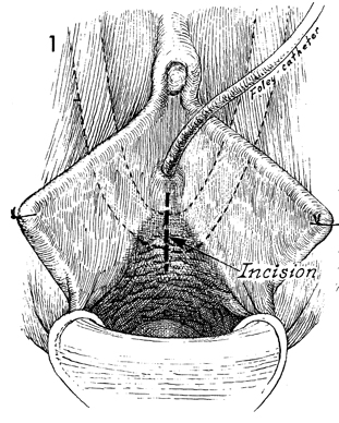
The patient is placed in the dorsal lithotomy position with the vulva and vagina prepped and draped. A Foley catheter is placed in the bladder. The exact location of the fascia lata strap is located by palpation, and a 3-4 cm vaginal mucosa incision is made.

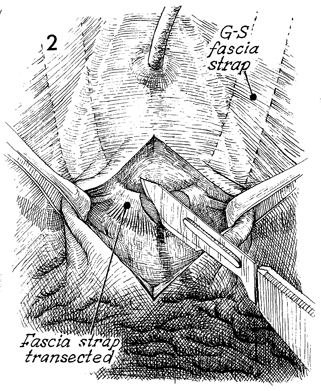
The vaginal mucosa is separated with Allis forceps. The strap is located and picked up with thumb forceps and is transected with a small knife. G-S identifies the Goebell-Stoeckel strap.

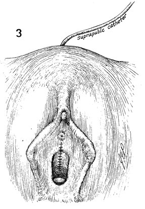
The edges of the vaginal mucosa are approximated in the midline with 3-0 synthetic absorbable suture. If necessary, a Foley catheter can be inserted transurethrally or suprapubically.


Copyright - all rights reserved / Clifford R. Wheeless, Jr., M.D. and Marcella L. Roenneburg, M.D.
All contents of this web site are copywrite protected.