Home / Site Map / Vulva and Introitus / Vagina and Urethra / Bladder and Ureter / Cervix / Uterus
Fallopian Tubes and Ovaries / Colon / Small Bowel / Abdominal Wall / Malignant Disease: Special Procedures

Malignant Disease:
Special Procedures

Staging of Gynecologic
Oncology Patients With
Exploratory Laparotomy

Subclavian Port-A-Cath 

Peritoneal Port-A-Cath

Application of Vaginal
Cylinders for Intracavitary
Radiation Therapy

Application of Uterine Afterloading Applicators
for Intracavitary Radiation Therapy  

Pelvic High-Dose Afterloader

Abdominal Injection of Chromic Phosphate
()

Supracolic Total Omentectomy

Omental Pedicle "J" Flap

Tube Gastrostomy

Total Vaginectomy

Radical Vulvectomy
With Bilateral Inguinal
Lymph Node Dissection

Reconstruction of the
Vulva With Gracilis Myocutaneous Flaps

Transverse Rectus
Abdominis Myocutaneous
Flap and Vertical Rectus
Abdominis Myocutaneous
Flap

Radical Wertheim
Hysterectomy With
Bilateral Pelvic Lymph
Node Dissection and With Extension of the Vagina

Anterior Exenteration

Posterior Exenteration

Total Pelvic Exenteration

Colonic "J" Pouch Rectal
Reservoir

Kock Pouch Continent Urostomy

Omental "J" Flap Neovagina

Ileocolic Continent Urostomy (Miami Pouch)

Construction of Neoanus
Gracilis Dynamic Anal
Myoplasty

Skin-Stretching System Versus Skin Grafting

Gastric Pelvic Flap for
Augmentation of Continent Urostomy or Neovagina

Control of Hemorrhage in Gynecologic Surgery

Repair of the Punctured
Vena Cava

Ligation of a Lacerated
Internal Iliac Vein and
Suturing of a Lacerated Common Iliac Artery

Hemorrhage Control in
Sacrospinous Ligament
Suspension of the Vagina

Presacral Space
Hemorrhage Control

What Not to Do in Case of Pelvic Hemorrhage

Packing for Hemorrhage
Control

Control of Hemorrhage
Associated With Abdominal Pregnancy

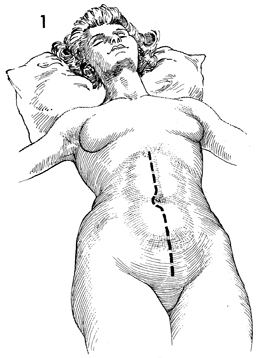
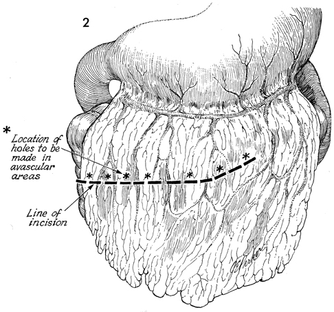
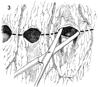
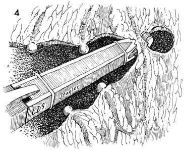
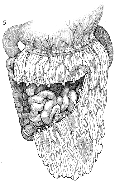
Omental Pedicle "J" Flap

An omental "J" flap provides (1) a nonirradiated vascular pedicle flap to cover intestinal anastomoses and (2) vesicovaginal-rectovaginal fistula repairs to form a lid on the inlet of the true pelvis after exenteration and to form a cylinder for a neovagina.

The purpose of this operation is to create a flap from the omentum by transecting the omentum from its attachments to the stomach, leaving enough branches of the left gastroepiploic vessels to provide an adequate blood supply for the flap.

Physiologic Changes. Irradiation produces obliterative endarteritis, ischemia, and fibrosis, all of which retard healing. By applying a vascular pedicle that has not been irradiated, the surgeon attempts to reverse some of the ischemia present in the irradiated tissue by promoting capillary and arterial ingrowth from the pedicle flap's blood supply. In addition, when the inlet to the true pelvis has been blocked by an omental lid, the small bowel is prevented from dropping into the denuded true pelvis after an extensive operation. Therefore, the possibility of intestinal obstruction and fistula formation is reduced. The omentum has a copious blood supply. Therefore, it is an excellent recipient of a skin graft for a neovagina.

Points of Caution. The short gastric vascular arcades to the omentum must be identified on the greater curvature of the stomach prior to initiating the procedure to ensure an adequate blood supply from the gastroepiploic artery remains for the proposed omental flap. The flap should be designed so the stomach is not pulled into the lower abdomen. The flap should not be placed on tension.

Technique

This operation is performed in conjunction with other radical pelvic surgery. Therefore, the appropriate incision for the initial procedure is adequate for the omental "J" flap. It is extremely difficult to perform the omental J flap through a transverse or Pfannenstiel incision, so a midline incision extended around the umbilicus is preferred.

The design of the flap prior to transecting the omentum is essential. A centimeter ruler and unfolded sponge are helpful in determining the appropriate length needed for the flap to reach the pelvis without tension. A check of the vascular arcades should be made to ensure that an ample blood supply is entering the base of the flap. Generally, the transection of the omentum is started at the hepatic flexure of the colon and proceeds from the patient's right to her left.

The omentum is opened in avascular areas with a small Kelly or Metzenbaum scissors. The vascular bridges between these openings can be doubly clamped with Kelly clamps, incised, and tied with 2-0 suture.

An alternative to Kelly clamps is the automatic LDS (linear dissecting) stapler (United States Surgical Corp.). This device clamps the vascular bridges between the openings in the omentum with the jaws of the stapler, applies two stainless steel clips, and activates a scalpel within the stapler to cut between the steel clips. It is a valuable, and time-saving device.

The omental flap is completed. It can be moved into the pelvis as a cover for a suture line or a pelvic lid.


Copyright - all rights reserved / Clifford R. Wheeless, Jr., M.D. and Marcella L. Roenneburg, M.D.
All contents of this web site are copywrite protected.